


 VIP会员


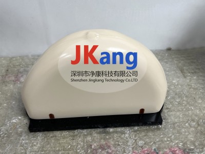
Cirs 011a mammogram model, cirs 011a mammogram x-ray tissue equivalence model recommended by the american cancer society and the radiology academy for mammography quality assessment
A sophisticated quality assurance tool for today's advanced imaging system。
Cirs 011a mammograms have been validated in modelling techniques that allow tissue to be equated and authentic models to be used for mammography quality assurance。
The resin material imitates the photon decay coefficient of a series of breast tissues. The average composition of the imitated human breast is based on individual elements of fat and gland tissue reported by hammerstein。
Decomposition factors are calculated using jhubbell's “mixed rules” and photon mass decay and energy absorption factor tables。
Cirs 011a mammograms contain targets aimed at testing thresholds for new generation mammography machines. 4. 5 cm thick and simulated mean gland tissue composition。
The purpose is to test the performance of any mammogram system. The matter in the model simulates calcification, fibre calcification in catheters and tumor blocks. The range of objects to be tested in the body range should range from objects that should be visible on any system to objects that are difficult to resolve on the best mammography system。
Cirs 011a mammogram, cirs 011a mammogram x-ray tissue technical parameters:
1. Line to details 1) 20 lp/mm 2 group horizontally and vertically
2. Calcium carbonate stoves (2) 0. 13), 0. 165 (4) 0. 196 (5) 0. 23 (6) 0. 275 (7) 0. 4 (8) 0. 236 (10) 0. 165 (11) 0. 196 (13) 0. 165
3. Wedge block (1cm) 14) 100% gland tissue 15) 70% gland tissue 17) 30% gland tissue 18) 100% fat tissue
4. Niron fibres (19) 1. 25 mm 20) 0. 83 mm 21 0. 71 mm 22) 0. 53 mm 23) 0. 3mm
5. Hemispheric mass (75% gland, 25% fat) 24) thickness 4. 76 mm 25) thickness 3. 16 mm 26) thickness 2. 38 mm 27) thickness 1. 98 mm 28) thickness 1. 59 mm 29) thickness 1. 19 mm 30) thickness 0. 9 mm
6. Photodensity 31) reference area
7. Lineline boundary 32) location details