

Eastern zidct operations manual
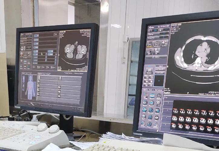
Read and understand this operating manual carefully before using toshiba ct equipment. This manual will guide you in completing the start-up, operation, day-to-day maintenance and security of the equipment. The following is a detailed guide to use. Equipment start-up and preparation 1. Equipment check, prior to operation, the appearance and functionality of the equipment. Ensure that the equipment power lines are robust and all attachments are complete, including scanner beds, consoles and necessary cables. 2. Power is activated by pressing the power button on the equipment, pending the start of the equipment. Normally, the start-up of the device takes a few minutes and the start-up progress is shown on the screen. After startup, enter the main interface. 3. User login using your username and password login system. Make sure you have proper permission to operate. If you have a login problem, contact the system administrator. Scan operation 1. Patients are prepared to ensure that they have made the necessary preparations as instructed before entering the scanning room. For example, some scans may require patient fasting or specific medication. Sets the scanning parameters in the main interface, selects the " scan " option, and then sets the scanning parameters as required. These parameters include the type of scan (e. G., smoothing, enhanced scanning), scanned range, layer thickness and spacing. 3. Patient orientation guides the patient to lie on a scanned bed and ensure that the patient is in the right place. Initial positioning is performed using a bedside x-ray machine to ensure the accuracy of the scanning range. When the scan is initiated to confirm all settings and patient locations, press the “start scanning” button. The equipment will begin data collection. During the scanning process, the patient is ensured to remain static and to avoid hypocrisy. 5. When the scan is completed, the device will alert you. At this point, the patient may be instructed to leave the scanned bed. Ensure that data are saved before seeing the results of the scan. The system automatically saves data when data processing and viewing 1. Data preservation scanning is completed. You can find these data in the specified folder. Ensure data preservation is complete and intact. Image viewing of data to be saved using the device's own image viewing software. Software usually provides multiple modes of view, such as two-dimensional and three-dimensional film models. Select the appropriate view mode as needed. 3. Image processing can be edited using tools provided by software if it is necessary to process the images. For example, comparisons, brightness, thin layer reconstruction, etc. Can be adjusted. Daily maintenance 1. Cleaning equipment is used daily to wipe the surface of the equipment with clean cloth and remove dust and stains. For scanning beds and cables, cleaning is done using a special cleaning agent. 2. Periodic inspection of the equipment's functionality, such as smooth movement of the scanner bed and strong cable connection. If any problems are identified, contact the maintenance staff in a timely manner. 3. Software updates regularly check and update equipment software to ensure that the equipment is operational in its latest version. Software upgrades can often be done through networks to ensure that network connections are stable. Safety precautions 1. Patients are safely scanned to ensure a safe environment. Avoiding exposure of patients to any metal object to prevent the risk of electroshock. 2. Operators shall wear appropriate protective equipment such as protective glasses and gloves. Direct contact with scanned areas of patients is avoided to reduce radiation exposure. 3. Emergency situations are processed in the course of scanning and, in the event of an emergency (if the patient is not well or equipment is malfunctioning), the emergency stop button is immediately pressed and processed in accordance with the contingency plan. Failed to initiate 1. The equipment cannot be activated and the power lines and power outlets are inspected. If the problem remains, contact the maintenance staff for inspection. If the scan fails, check if the scanning parameters are set correctly. Ensure that the patient is located correctly and re-scan. 3. If the image quality is poor, check the scanned parameter settings, such as layer thickness and layer spacing. You can try to adjust parameters or re-scan. A summary of the operations manual detailing the start-up, operation, daily dimensions of the ct equipment