

The “cell-cell communication” (cell-cell community, ccc) represented by extraterrestrial (or extra-cellular cysts) and their components (e. G. Rna, lipids and proteins), linear particle transfer (e. G., tunnel nanoconductor), formulation-receptor (e. G., membrane, genocidal, and membrane receptor) has been the subject of concern for many research teams, as well as for natural projects and high-profile articles in the country over the years. Today we're going to take an inventory of these three ways of working。
I. Inter-cellular communication with the guidance of the endocrine body
The excretion is a nanocystical cytological cytology carrying biological molecules such as proteins, rnas, which regulates their functions by integrating or internalizing receptors。
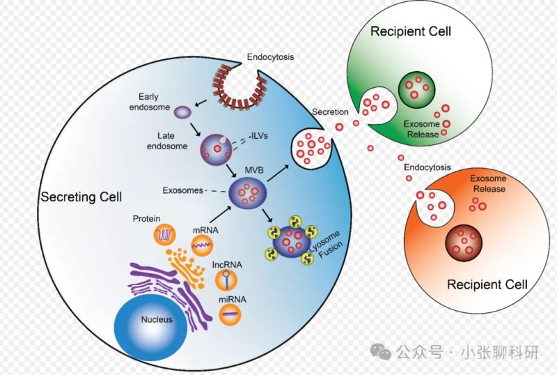
1) methods of separation, identification and representation of the excreta
Traditional methods such as hypervelocity centrifugals, density gradients, filtration, overfiltration, etc. Are commonly used for the separation of the excretion, and new technologies such as microballs and microflow control chips are also available. Protein markers such as cd9, cd63, cd81, etc., are commonly used for the testing of excreta. The manifestations include morphological and particle distributions, which can be used using a penetrating electron microscope (tem), nanoparticle tracking analysis (nta), etc。
2) inter-cell transfer method for trace of the excretion
Tracement methods include fluorescent dye markings, such as pkh26, pkh67, which can be directly associated with the excrete lipid double membrane; nucleic acid markings, which mark the excrete nucleic acid as fluorescent or radioactive probes; protein tags, which mark the excrete surface proteins, such as gfp; and magnetic nanoparticles tags, which can also be used to track through mri imaging。
3) organizational analysis of the components of the endocrine system
The main components of octology analysis include protein, transfer and lipid. Proteomics analyse the composition of proteins in the excrete through mass spectrometry techniques; transcribing spectrometry studies the rna species and expression levels in the excrete using high flux sequencing techniques; and lipids use mass spectrometry to analyse the composition of lipids in the excrete。
4) cultivation of endocrine and receptor cells
The purified excretion is added to the receptor cell culture fluid, and the effects of the excretion on the receptor cell are studied by observing the reaction and appearance of the receptor cell. In addition, co-production can be followed by the marking of the excretion and real-time monitoring of its dynamics in receptor cells。
Lineh particles can be transferred from one cell to another by means of nanopipes (tnts) in the tunnel, by means of extracellular bubbles or by direct cell release, to support the metabolic needs of receptor cells。
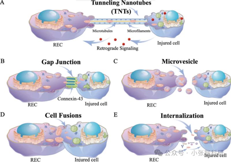
1) line particle separation and identification
Line particle separation is usually carried out by a differential centrifuge method, which removes cellular fragments and nucleus first at a low pace, followed by high-speed centrifuge sediment particles. Validation methods include penetrating electron microscopes (tem) to observe their bipolar membrane structure, as well as western blot testing through specific antibodies (e. G. Anti-tom20 antibodies)。
2) post-separation pellets with cell culture
The separated line particles are added to the receptor cell culture fluid to observe whether the line particles are ingestion through fluorescent microscopes or current cytology. Chemically induced feed cell line particles can also be developed in conjunction with receptor cells after depletion, comparing the differences between receptor cells。
3) the marking and trace of linear particles under the cell co-culture system
Fluorescent dyes (e. G., mitotracker) or fluorescent proteins (e. G., gfp) are used to mark line particles of feed cells, which are then nurtured with receptors. The distribution of particles in receptor cells through fluorescent microscopes or co-focused microscopes, resulting in the transfer of trace line particles。
4) single-cell sequence combined with merci analysis
When data are obtained for the transfer group of co-breeding cancer cells and t cells, the merci algorithm is used to identify the mutation of the mitsnvs and to estimate the relative abundance of the exterior ray particles in the cancer cell. By comparing mutation frequencies, t-cell-specific mutations are identified, classifying cells as potential linear particle providers and receptors. By calculating the scores of dna and rna rankings, receptors and feed cells of line particles are projected。
When the formulation is combined with the receptor, it induces the receptor to change, activates the downstream signal route and regulates the cell function。
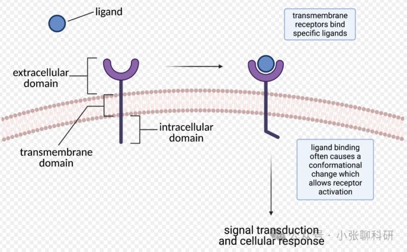
1) disruptive antibodies disrupt receptor activity
Use of a barrier-resistant antibody for a specific area of the receptor, which can bind the receptor specifically to prevent the combination of the formulation and the receptor, thereby inhibiting the receptor's activation. By detecting the activation of downstream signals (e. G., protein phosphorus acidization levels), the disruption effects are assessed and used to study the function of receptor activation and signal transfer mechanisms。
2) moderate antibodies reduced formulation concentrations
Using neutral antibodies combined with the specific properties of formulations, antibody-composite compounds are formed, thus reducing the concentration of ionomers. By detecting the reaction of cells to formulations (e. G. Cell proliferation, cell-factor genocrine, etc.), the moderate effects are assessed and used to study the role and control mechanisms of the formulations in the physiological process。
3) formulation - dyeing and co-location with receptor under tissue and cell culture
In co-production systems, formulations and receptors are dyeed with fluorescent marked antibodies. The co-location is analysed by observing the fluorescent signals of both by fluorescent microscopes or co-focus microscopes. The co-positioning analysis uses software to calculate a co-positioning factor to determine whether the ingredient and the receptor are concentrated in the same cell area, thus insulating their interaction under physiological conditions。
4) immuno-sedimentation validation formulations - combined with receptor
After cell cracking, specific antibodies are added to the receptor, and the antibodies are hatched to bind the receptor. Add protein a/g glycerine, which combines antibody with beads to settle receptor protein complexes. Finally, the presence of formulation proteins in sediments is tested through western blot, and if the formulations are detected, the combination of the two in the cells is shown。
5) surface plasma resonance test formulation-receptor combination strength
The receptor is attached to the cpr chip surface, and the formulation passes through the chip surface in the flow phase. The spr technology allows real-time monitoring of refractive rate changes in the combination of formulations and receptors, thereby obtaining kinetic parameters such as combined proximate strength (kd), combined rate constant (ka) and deviation rate constant (kd) for quantitative analysis of their combined strength。
6) analysis of rectification of receptors in combination with receptors
The receptor is detected by means of techniques such as round two-colour spectra (cd) or mri (nmr). Cd can analyse secondary structural changes and nmr can provide more detailed three-dimensional structural information. In addition, fluorescent resonance energy transfer (fret) technology can be used to indirectly reflect image change by detecting internal distance changes in receptors through fluorescent markers。